
Figure 4.
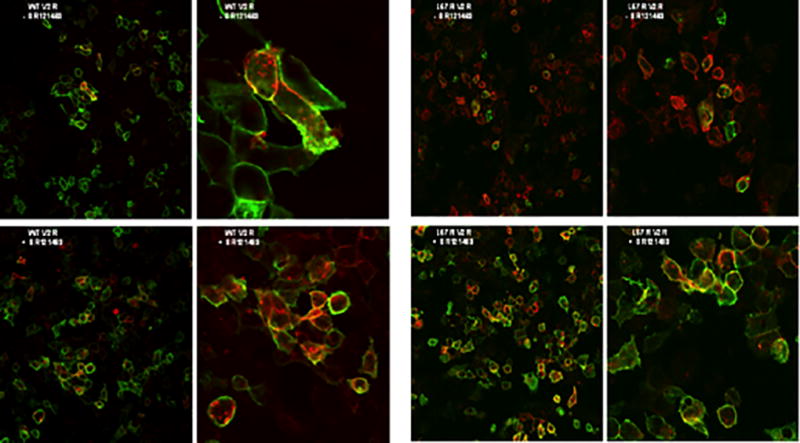
Membrane expression of V2R. Nonpermeabilized HEK 293 cells expressing myc-tagged WT or L57R V2R and farnesylated mCherry were incubated with and without 10−6 M SR121463 for 48h and imaged by fluorescence microscopy. Red fluorescence identifies the cell membrane of all transfected cells. Green fluorescence identifies the WT or mutant receptor when it is present at the cell membrane.