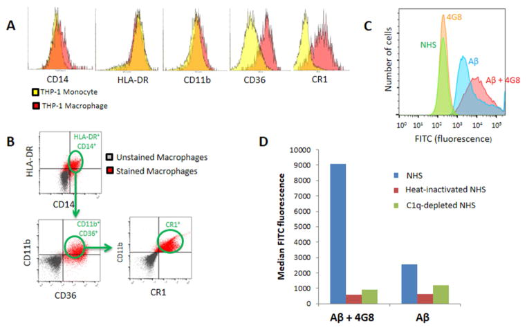
Fig. 4. Macrophage capture of Aβ and Aβ ICs.
A) Flow cytometry characterization of THP-1 monocytes and their differentiation to THP-1-derived macrophages revealed a transition to increased expression of inflammatory markers such as CD36, a mediator of macrophage phagocytosis [25], and, especially, CR1, the macrophage receptor for the major complement opsonins in primates [34,35]. B) To insure assessment of the macrophage-specific phenotype in subsequent experiments, all cells were gated for criterion expression of five different macrophage markers, CD14, HLA-DR, CD11B, CD36, and CR1. C) Fluorescence intensity distributions for samples pre-incubated with NHS. Distributions designated NHS and 4G8 represent fluorescence of the macrophage markers on which the cells were gated. Fluorescence distributions for gated, FITC-labeled Aβ and Aβ IC samples appear in the right-hand portion of the graph. A total of 100,000 cells were evaluated for each sample. D) Median fluorescence intensity of Aβ captured by THP-1-derived macrophages increased nearly 4-fold for Aβ ICs compared to Aβ alone. Only background readings were obtained for the cells when heat-inactivated NHS or C1q-depleted serum were substituted for NHS as a complement source.

