
Figure 4. MALAT1 regulates neointima formation during vascular injury.
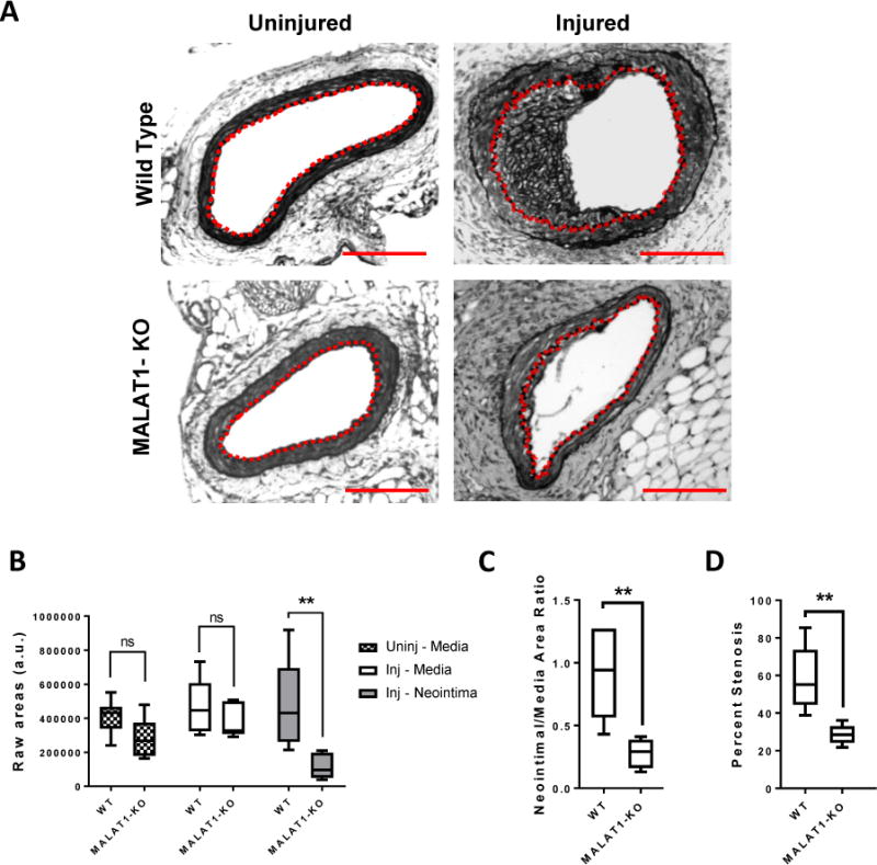
(A) Cross-sections of injured and uninjured (contralateral) femoral arteries of wild-type (WT) and MALAT1-knockout (KO) adult mice were stained for elastin to visualize the internal and external elastic lamina. The dashed red line delineates the internal elastic lamina, which circumscribes the neointima. Scale bar = 100μm. (B) Areas of the media and neointima from injured femoral arteries (inj – media, inj – neointima) and uninjured femoral arteries (uninj – media) of WT and KO mice. (C) Neointimal-Media area ratios of injured WT and KO femoral arteries. (D) Percent stenosis was calculated by dividing the neointimal area by the area circumscribed by the internal elastic lamina × 100%. (n = 7 WT, n = 5 MALAT1-KO, ** p<0.005, ns = not significant, Mann-Whitney Test).