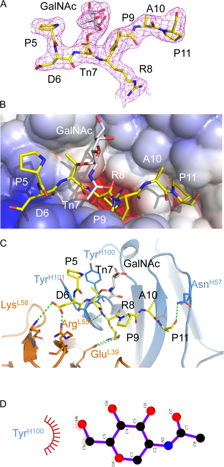
Fig. 3.
X-ray structure of AR20.5 in complex with MUC1 glycopeptide (APDTnRPAP). (A) 2Fo-Fc electron density map of MUC1 glycopeptide bound to AR20.5. The N-terminal residue A4 was disordered in the structure. There is clear electron density for the GalNac carbohydrate (gray). (B) Electrostatic surface of AR20.5 combining site. The MUC1 glycopeptide antigen binds in a surface groove. Residue R8 of MUC1 binds in a deep negatively charged pocket. (C) Binding interactions of MUC1 glycopeptide (yellow) with AR20.5. Binding is mediated by a series of electrostatic interactions and hydrogen bonds to both the heavy chain (blue) and the light chain (orange). The GalNAc carbohydrate (gray) makes no specific polar contacts with the antibody. (D) Nonpolar interactions between GalNAc and Tyr100H figure generated with LigPlot+ (Laskowski and Swindells 2011). This figure is available in black and white in print and in color at Glycobiology online.

