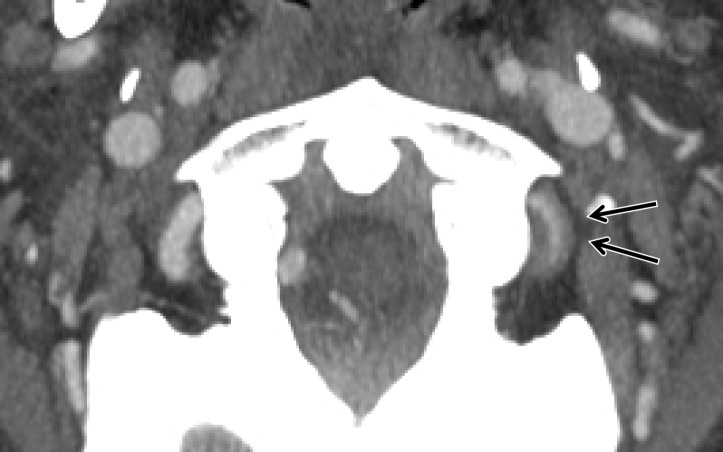
Figures 10a.
Intramural hematoma and intraluminal thrombus. Axial (a) and sagittal (b) CT angiographic images at the C1 level demonstrate left V3 segment vertebral artery wall thickening and luminal narrowing (>25%) (black arrows), representing intramural hematoma, as well as a focal luminal thrombus (white arrow in b), compatible with grade II injury.

