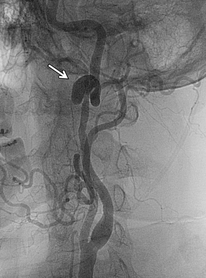
Figure 14c.

Pseudoaneurysm in a patient with minimal trauma but who had a family history of dissections. Axial (a) and sagittal (b) MIP CT angiographic images and lateral neck digital subtraction angiographic image following a left common carotid artery injection (c) show a large saccular outpouching of contrast material from the left ICA (arrow), representing a pseudoaneurysm and compatible with grade III injury.
