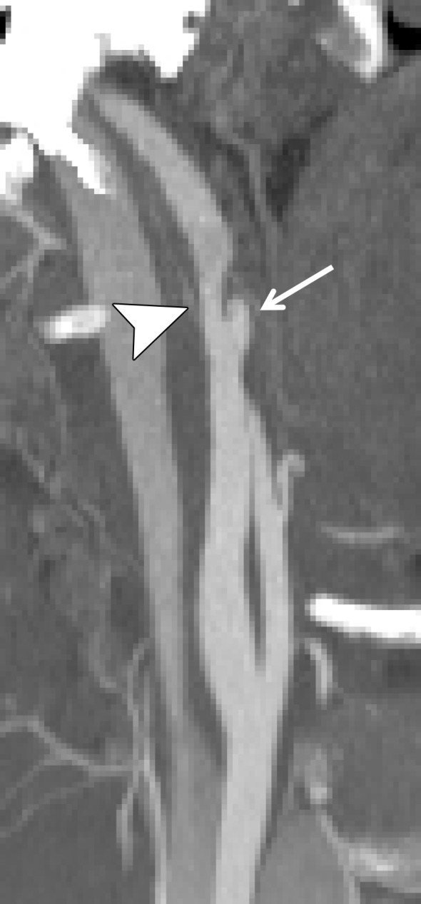
Figure 15a.

Pseudoaneurysm with enlargement at follow-up imaging in a polytrauma patient with cervical spine fractures. (a) Sagittal oblique MIP CT angiogram shows a pseudoaneurysm of the right cervical ICA (arrow), representing a grade III injury. The pseudoaneurysm and associated mural hematoma narrow the adjacent lumen (arrowhead). (b) Sagittal oblique CT angiogram obtained 7 days later shows slight interval enlargement of the pseudoaneurysm (arrow).
