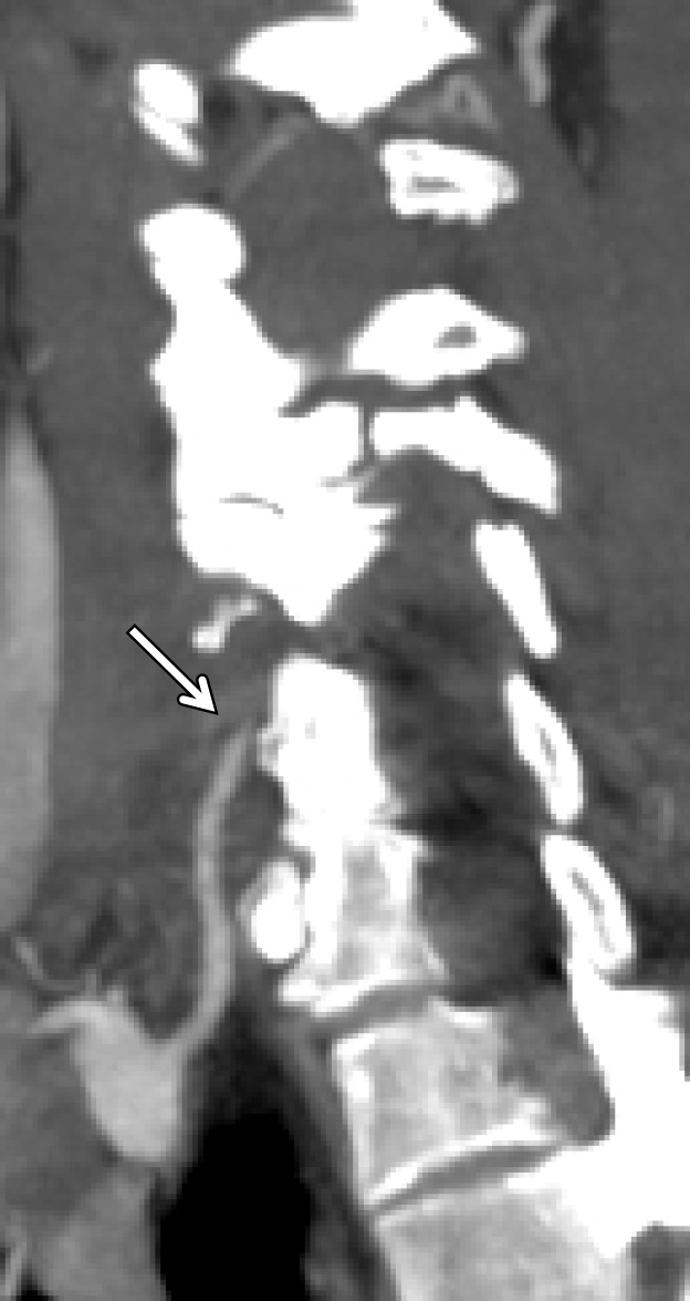
Figure 17a.
Vertebral artery occlusion. (a, b) Sagittal oblique (a) and sagittal (b) CT angiographic images show occlusion of the proximal right vertebral artery (arrow in a), with reconstitution distally at the C3–C4 vertebral level (white arrow in b), compatible with a grade IV injury. Fracture of the C5 articular facet (black arrow in b) is also seen. (c, d) Axial CT angiographic (c) and axial T2-weighted MR (d) images demonstrate occlusion (arrow in c) and loss of right vertebral artery flow void (arrowhead in d).

