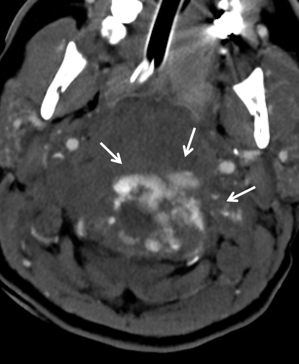
Figure 18a.
Vertebral artery transection in a polytrauma patient with atlanto-occipital dissociation. (a) Axial CT image shows active extravasation of contrast material within a large hematoma at the widened craniocervical junction (arrows). (b, c) Axial (b) and coronal (c) MIP images from CT angiography demonstrate hemorrhage from the transected left V3–V4 segment vertebral artery at the C1 level (arrows), representing grade V injury.

