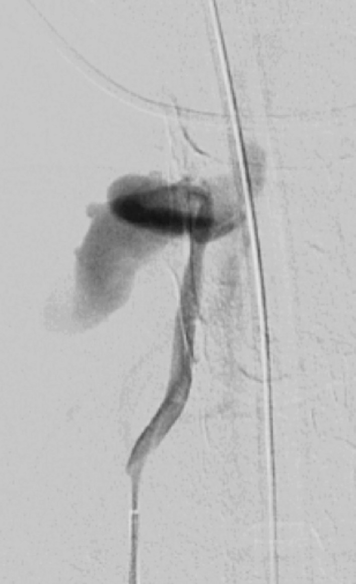
Figure 19d.

Vertebral artery transection and AVF in a polytrauma patient with C4–C5 fracture dislocation. (a) Coronal MIP image from CT angiography shows active extravasation with a hematoma (arrow) centered in the right C4 transverse foramen at the level of the fracture-dislocation. (b) Right vertebral artery injection catheter angiogram shows active extravasation, representing transection of the right vertebral artery (arrow), compatible with grade V injury. (c) Left vertebral artery angiogram shows retrograde flow into the right vertebral artery, to the level of the transection (arrow). (d–f) Right vertebral artery injection digital subtraction angiographic images in three subsequent phases show early venous drainage, compatible with AVF.
