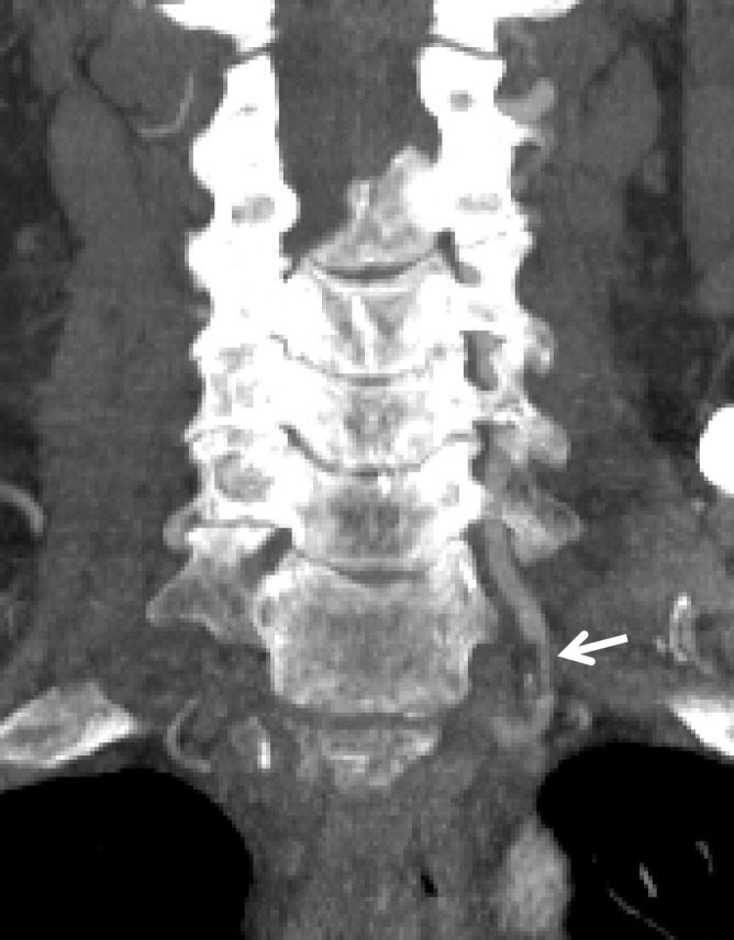
Figure 20a.
Atherosclerosis mimicking BCVI in a polytrauma patient at high risk for BCVI. (a) Coronal MIP image from CT angiography shows a mural lesion causing narrowing of the proximal left vertebral artery (arrow). Associated calcification is present, compatible with atherosclerotic plaque. (b) Axial CT angiographic image at the C1 level shows a calcified atherosclerotic plaque of the right V3 vertebral artery, causing narrowing and wall thickening (arrow), mimicking a stenotic BCVI (ie, intramural hematoma).

