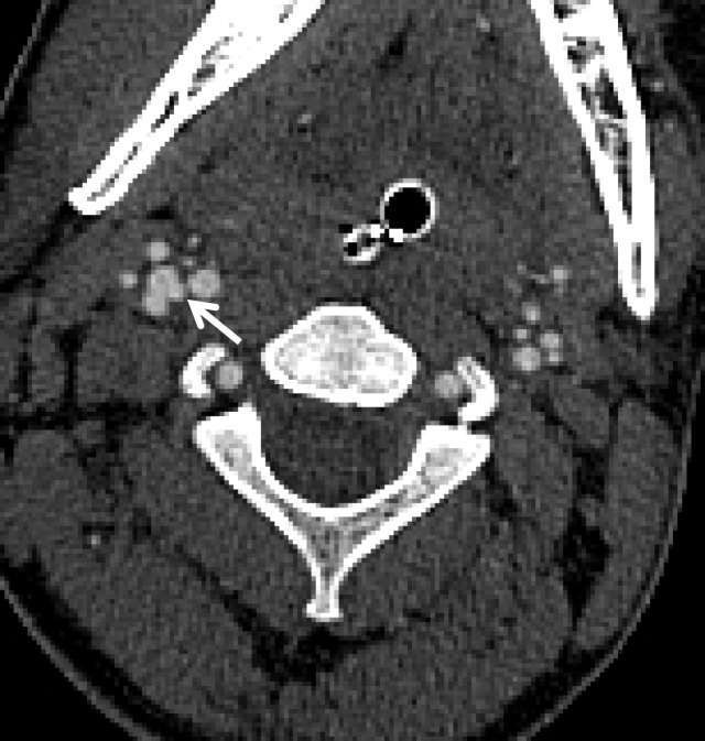
Figure 21a.
BCVI involving coiled or looped vascular segments. Coiled or looped vascular segments make evaluation for luminal and wall abnormalities difficult. Axial (a) and coronal (b) MIP images from CT angiography show a small medially directed pseudoaneurysm (arrow), which is easily ignored as it subtly projects from the coiled segment.

