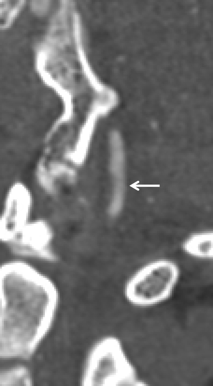
Figure 25a.

Worsening of a low-grade injury in a polytrauma patient with right occipital condyle fracture. (a) Oblique CT angiogram of the right vertebral artery at the time of injury shows an intramural hematoma (arrow) with 25%–50% narrowing, representing grade I–II injury. (b, c) Oblique (b) and frontal projection 3D-rendered (c) images from CT angiography obtained 7 days later show development of a small pseudoaneurysm (arrow), which represents progression to a grade III injury.
