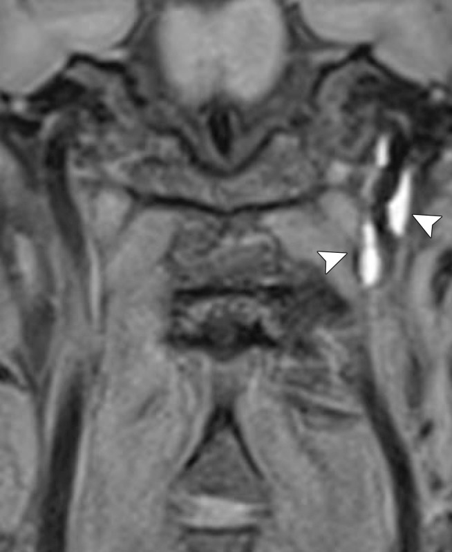
Figure 26e.
Vessel wall imaging of left ICA dissection. (a, b) Axial (a) and oblique (b) images from initial CT angiography show a high-grade stenosis, with a thin string of contrast material opacification (arrow), proximal to the skull base. Vessel wall MR imaging was performed 5 weeks later. (c) Three-dimensional time-of-flight MIP image shows improvement of the stenosis, with only minimal remaining luminal narrowing (arrow). (d, e) Axial (d) and coronal (e) black-blood fat-saturated T1-weighted MR images show persistent hyperintensity (arrowheads) within a thickened arterial wall, compatible with a subacute intramural hematoma. Note that, unlike the luminal time-of-flight MR angiogram, the vessel wall MR imaging sequences delineate the extent of true vessel wall disease.

