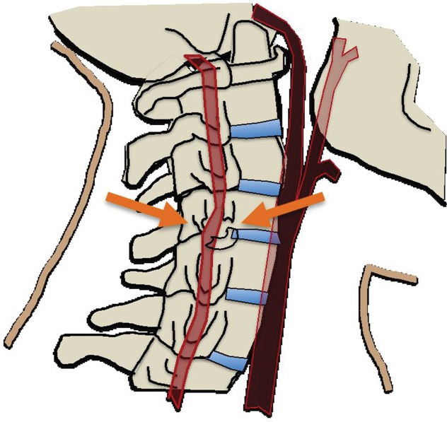
Figure 2d.
(a) Longitudinal stretching and/or twisting of the vessel. Illustration depicts hyperextension and contralateral rotation, with stretching of the ICA over the C1–C3 transverse processes (red dashed arrows). Blue arrows in a–c = direction of movement. (b) Vessel impingement. Illustration depicts compression of the ICA between the mandible and spine (yellow arrows), which may result from hyperflexion or displaced mandible fracture. Impingement may also result from skull base or cervical spine fracture. Likewise, a blow to the neck or strangulation may directly compress or impinge a vessel against an adjacent structure. (c) Stretching of the vessel. Illustration depicts stretching of the vertebral artery between subluxated or dislocated bone structures (red dashed arrows), which may occur in cases of cervical fracture, dislocation, or ligamentous injury. (d) Direct laceration. Illustration depicts a fracture with direct injury to the vertebral artery by an adjacent bone fragment (orange arrows). This is especially relevant in cases of vertebral transverse foramen or carotid canal fracture.

