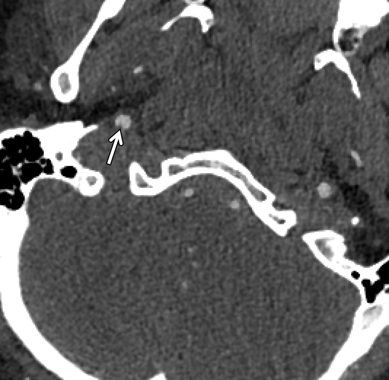
Figure 4a.
BCVI in a young patient who underwent strangulation. (a, b) Axial (a) and sagittal (b) CT angiographic images show focal irregularity of the right ICA with wall thickening, causing mild narrowing (arrows), proximal to the skull base. DSA was performed to further define the injury. (c) Digital subtraction angiographic image shows a focal filling defect (arrow) at the site of injury, compatible with intraluminal thrombus versus intramural hematoma.

