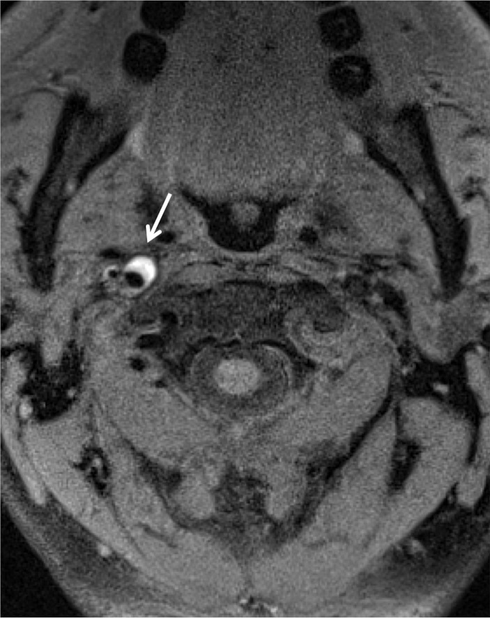
Figure 5a.
ICA dissection. (a) Axial fat-saturated T1-weighted MR image shows a hyperintense crescent (arrow) about the right ICA flow void, representing the false lumen. (b) Surface-rendered three-dimensional (3D) reconstruction of DSA data following right ICA injection shows irregularity and narrowing of the distal cervical right ICA (arrows).

