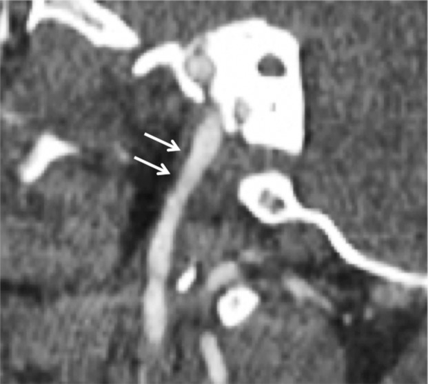
Figure 8a.
Minimal intimal injury in a trauma patient. (a) Coronal CT angiogram shows what appears to be minimal luminal irregularity (arrows), compatible with a grade I injury. (b, c) Axial (b) and coronal (c) fat-saturated T1-weighted MR images show normal signal intensity in the vessel walls (arrows). Lack of intramural hematoma or wall thickening suggests that the CT angiographic findings (a) likely represent artifact or chronic irregularity.

