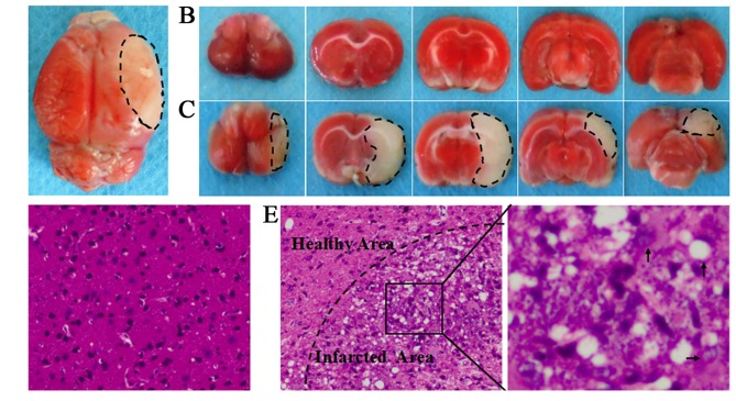
Figure 2.
(A) The complete brain of middle cerebral artery occlusion (MCAO) models. (B) Serial coronal slices of sham-operated rats. (C) Consecutive coronal slices derived from MCAO models. 2,3,5-Triphenyltetrazolium chloride (TTC) staining showed red healthy zones and pale infarcted regions; Cerebral infarct volume was ~32.1±1.8% in saline group. (D) H&E staining of sham-operated animals revealed intact brain tissues, with uniform, round or oval neurons and pink cytoplasm with abundant and clear nucleus. No inflammatory cells were detected. (E) H&E staining of MCAO models revealed lesions in the brain tissues, with diminished numbers of neurons and chaotic neuronal configuration. The cytoplasm was pale, eosinophilic and vacuolated. In addition, necrotic neurons with karyopyknosis, cellular gaps, debris and inflammatory cell infiltration were seen (×200).

