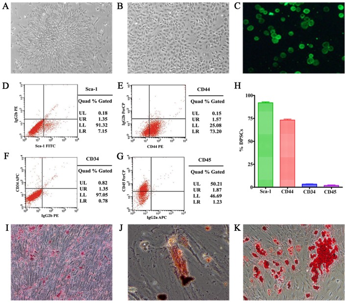
Figure 3.
(A) P0 of dental pulp stem cells (DPSCs), showing plastic adherence and colony formation. (B) P3 of DPSCs with fibroblast-like morphology. (C) PKH67-labeled DPSCs emit green fluorescence under a fluorescent microscope at 488 nm (×40). (D-G) Fluorescence-activated cell sorting (FACS) of DPSCs. (H) Surface marker expression of the isolated DPSCs: 91.32% expressed Sca-1; 73.20% expressed CD44; 1.35% expressed CD34; and 1.23% expressed CD45. (I-K) Multilineage differentiation of DPSC: mineralized nodules, calcium deposits and fat droplets (×100).

