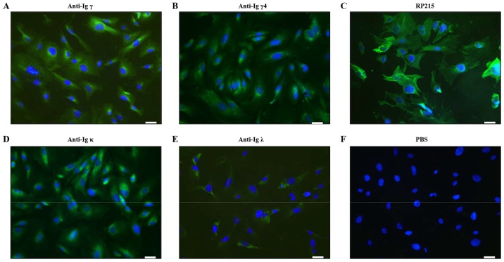
Figure 1.
IgG was detected in human podocytes by immunofluorescence staining. (A) Anti-Igγ, (B) anti-Igγ4, (C) RP215, (D) anti-Igκ and (E) anti-Igλ staining; (F) PBS was used as a negative control. Igs were immunostained (green) and nuclei were stained with DAPI (blue). Scale bar, 50 µm. IgG, immunoglobulin G; Igγ, IgG heavy chain.

