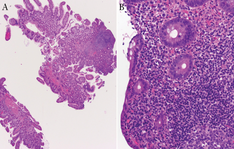
Figure 2.
Biopsy of the ileal pouch from patient B. (A) Under low power, the small intestinal type mucosa shows focal villous blunting and a lymphoid follicle (original magnification x 40). (B) High power view shows focally decreased mucin and neutrophilic infiltration in the lamina propria and epithelium (original magnification x 400).

