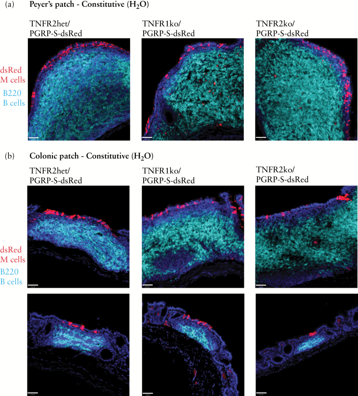
Figure 2.
Constitutive Peyer’s patch and colonic patch M cell development is intact in TNFR1 and TNFR2 knockout mice. a. Expression of PGRP-S-dsRed transgene reporter [red] in M cells, with immunostaining for B cells [cyan] shows normal development and organisation of Peyer’s patches in TNFR1 and TNFR2 knockout mice. Scale bar: 40 µm. b. Constitutive colonic patch B cell follicle [cyan] organisation and associated M cells [red] are intact in TNFR1 and TNFR2 knockout mice. Scale bar: 40 µm.

