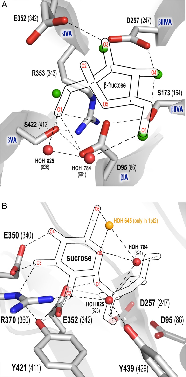
Fig. 3.
Network of contacts within the −1 (A) and +1 (B) subsites of B. subtilis levansucrase (1oyg, numbering in brackets). Fructose (A), glucose (B) and structural water molecules were superimposed from 1pt2. Dashed lines: contacts within 2.5–3.5 Å. β-sheets I–VA are labeled in blue font. (A) Contacts of fructose with the catalytic amino acids, S173, S422 and R353; (B) network of contacts of R353 within the active site connecting the β-sheets. This figure is available in black and white in print and in color at Glycobiology online.

