Figure 5.
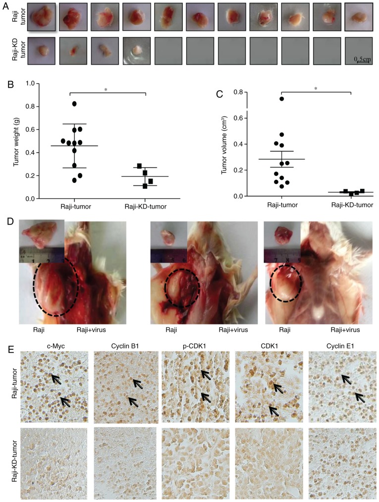
Knockdown of c-Myc suppresses the tumorigenesis of Raji cells in a SCID mouse xenograft model. (A-C) At 30 days after injection of 1×10⁷ Raji or Raji-KD cells into the axilla, tumors were harvested. Tumor formation at the site of injection was monitored. (A) Representative images of tumors formed in the mice. (B) Tumor weight in the two groups. The weight of Raji-KD tumors was lower than that of Raji tumors. (C) Tumor volume in the two groups. The volumes of Raji-KD tumors were smaller than those of Raji tumors. Values are expressed as the mean ± standard error of the mean. *P<0.05. (D) Representative images of tumors grown in the animals at 45 days after injection of 2×10⁶ Raji cells into the bilateral axillary sites of the hind legs (circles with broken lines). On the first two days after inoculation of Raji cells, mice were injected in to the axilla with c-Myc shRNA retrovirus on the right or with the pseudotyped retrovirus on the left. Magnified images display the respective tumors after dissection. The tumor formation of Raji cells was inhibited by injection of c-Myc shRNA retrovirus. (E) Immunohistochemical staining of Raji (upper panels) and Raji-KD (lower panels) xenograft tumor tissues [from (A)] for c-Myc, CDK1, p-CDK1, cyclin B1 and cyclin E1 (magnification, ×200). Immunopositivity was indicated by a brown stain (arrows). shRNA, small hairpin RNA; p-CDK1, phosphorylated cyclin D kinase 1; Raji-KD, Raji cells with c-Myc knockdown.

