
Figure 3.
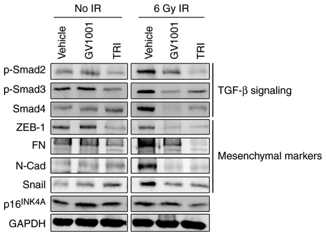
GV1001 suppresses TGF-β signaling and EMT in NHOKs exposed to IR. NHOKs were exposed to 6 Gy IR and maintained in culture for 10 days in the presence of GV1001 (1 µM) or TRI (1 µM). Western blot analysis was performed for TGF-β signaling molecules, p-Smad2/3 and Smad4, and TGF-β target mesenchymal markers, ZEB1, FN, N-Cad and Snail. GAPDH was used as a loading control. NHOKs, normal human oral keratinocytes; IR, ionizing radiation; TGF-β, transforming growth factor-β; TRI, TGF-β receptor inhibitor; Smad, small mothers against decapentaplegic; p-, phosphorylated; ZEB1, zinc finger E-box binding homeobox 1; FN, fibronectin; N-Cad, N-Cadherin.