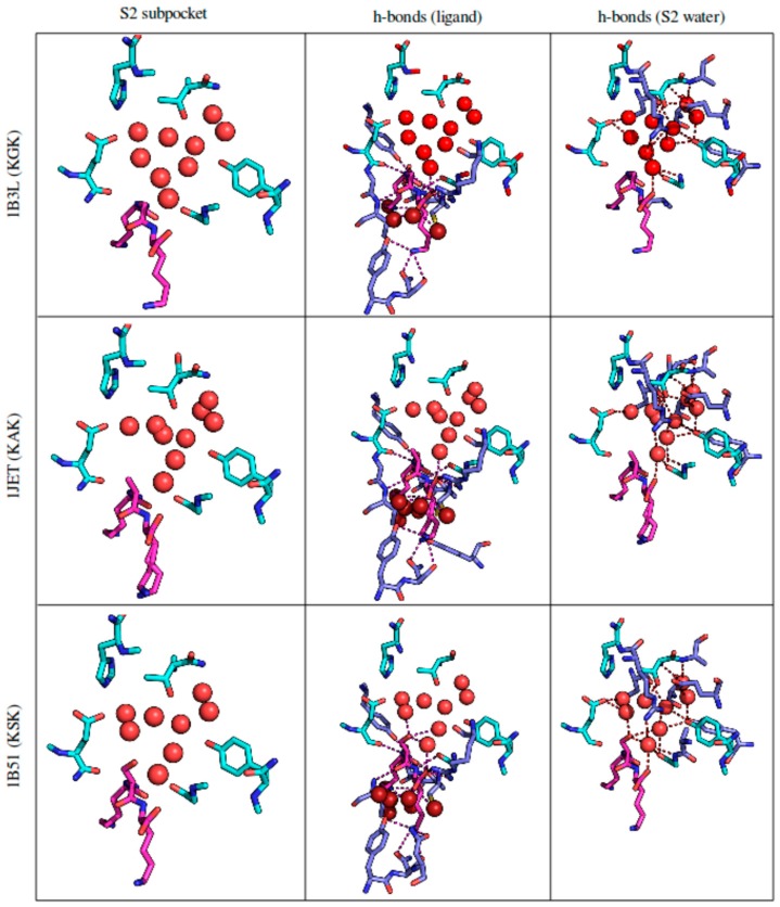
Figure 1.
The S2 binding pocket of OppA, taken from the respective crystal structures. Protein residues pointed out by reference [15] are colored in cyan. Protein residues other than these that make hydrogen bonds with either the ligand or S2 subpocket water are depicted in lavender blue. The tripeptide ligand and its hydrogen bonds are colored in magenta. Water molecules are depicted as spheres: Water in the S2 subpocket (pointed out by reference [15]) and its hydrogen bonds are colored bright red; water with bonds to the ligand is colored dark red. Hydrogen bonds were estimated by PyMol’s polar contacts function, which is based on DSSP. For details on individual water molecules see Figure S1 in the Supplementary Materials.

