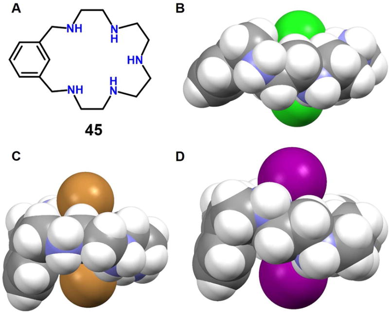
Figure 19. Polyaza metacyclophane anion receptors and its anionic dimer complexes.

(A) Molecular structure of receptor 45.
(B) Single crystal X-ray structure of the complex [Cl2]2−⊂ [H545]5+.
(C) Single crystal X-ray structure of the complex [Br2]2−⊂ [H545]5+.
(D) Single crystal X-ray structure of the complex [I2]2−⊂ [H545]5+.
All anions encapsulated within the cavity and the protonated 45 are shown in space-filling form. Inter-halide distances: (B) 3.51 Å, (C) 5.01 Å and (D) 5.30 Å. Color code: C = gray, N = blue, Cl = green, Br = brown and I = purple. The counter anions and solvent molecules outside the cavities are omitted for clarity.
