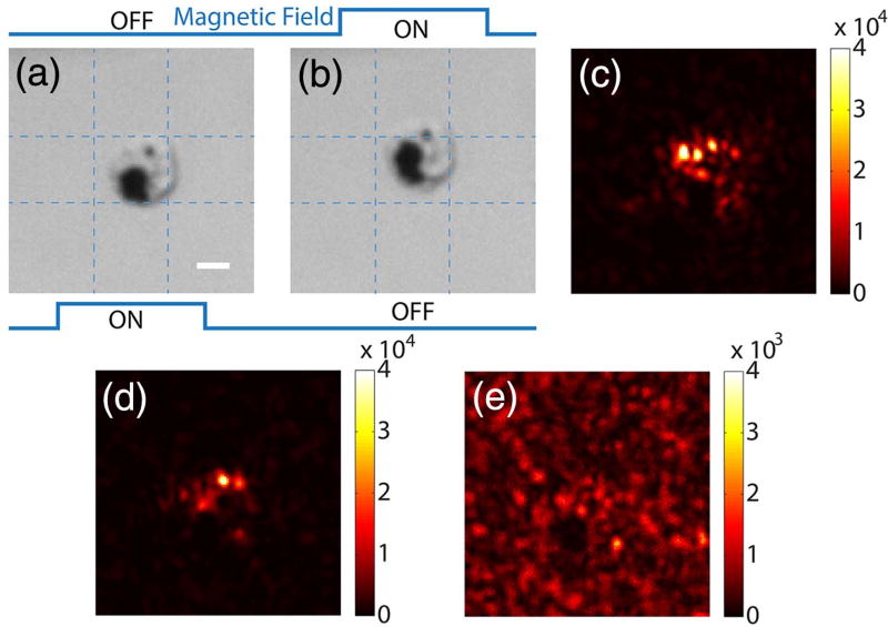
Fig. 4.
Focusing light onto a targeted cell that endocytosed magnetic particles of 453 nm diameter. Panels (a) and (b) show bright-field images of a cell under two magnetic fields. (c) Focus achieved by the field subtraction method. (d) Focus achieved by the frequency modulation method (fm = 25 Hz). (e) Control experiment: no focus was observed when the SLM pattern was circularly shifted by 10 × 10 pixels after obtaining the result in (d). Scale bar, 5 µm.

