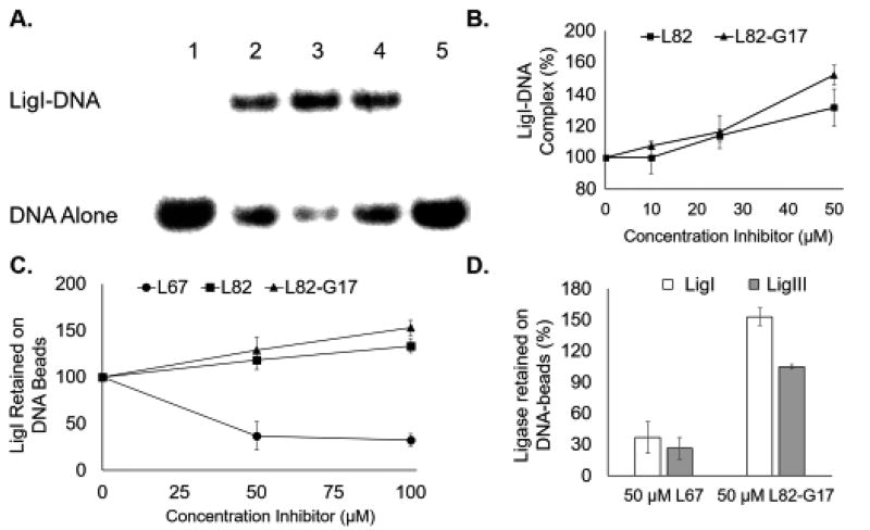
Figure 5. L82 and L82-G17 increase LigI binding to non-ligatable nicked DNA binding.
A. A representative gel showing the effect of the inhibitors, L67 (lane 5), L82 (lane 4) and L82-G17 (lane 3), at 100 µM and DMSO alone (lane 2) on DNA-protein complexes formed by LigI with non-ligatable nicked DNA. Lane 1, DNA substrate alone. The position of the DNA substrate (DNA alone) and the protein-DNA complex (LigI-DNA) are shown. B. Results of at least three independent EMSA assays are shown graphically with DNA-protein complex formation calculated as the ratio of bound and unbound DNA and expressed as percentage of the ratio obtained in reactions with LigI alone. L82 (closed squares) and L82-G17 (closed triangles). C. The effect of L82 and L82-G17 on the amount of labeled LigI retained by streptavidin beads liganded by biotinylated nicked DNA. Results of three independent assays are shown graphically and expressed as a percentage LigI retained in assays with DMSO alone. L82 (closed squares), L82-G17 (closed triangles) and L67 (closed circles). D. The effect of L67 and L82-G17 on the amount of labeled LigI (open squares) and LigIII (filled squares) retained by streptavidin beads liganded by biotinylated nicked DNA. Results of three independent assays are shown graphically and expressed as a percentage LigI/LigIII retained compared with assays with DMSO alone.

