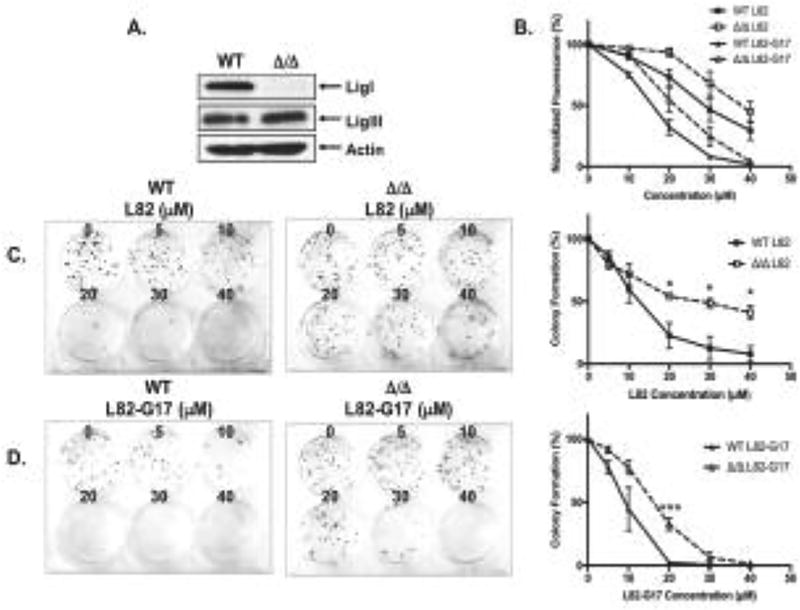
Figure 8. Cells lacking LigI are more resistant to L82 and L82-G17.
A. LigI, LigIII and β-actin proteins were detected in extracts of CH12F3 WT and CH12F3 Δ/Δ cells by immunoblotting. B. Effect of L82 (square) and L82-G17 (triangle) on the proliferation of CH12F3 WT (filled symbols) and CH12F3 Δ/Δ (empty symbols) cells was measured by the CyQUANT assay as described in Materials and Methods. Effect of L82 (C) and L82-G17 (D) on colony formation by CH12F3 WT and CH12F3 Δ/Δ cells. Data shown graphically are the mean ±SEM of three independent experiments and are expressed as a percentage of the values for the untreated cells. * p < 0.05 and *** p < 0.001 using the unpaired two-tailed Student test.

