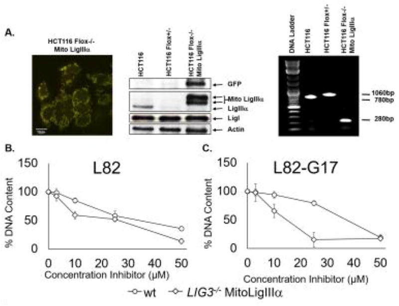
Figure 9. Cells lacking nuclear LigIIIα are more sensitive to L82 and L82-G17.
A. Fluorescent image of HCT116 Flox−/− expressing YFP-tagged mito LigIIIα. Scale bars, 10 µm (left panel). Immunoblots with extracts of HCT116, HCT116 Flox+/− and HCT116 Flox−/− Mito LigIIIα using antibodies against GFP, LigIII, LigI and β-actin. The positions of YFP-fusion protein (GFP), mito LigIIIα fused to GFP (Mito LigIIIα) and endogenous LigIIIα, LigI and β-actin are indicated (middle panel). Wild type and Floxed LIG3 alleles and the integrated cDNA encoding YFP-tagged Mito LigIIIα were detected in genomic DNA from HCT116, HCT116 Flox+/− and HCT116 Flox−/− Mito Ligase IIIα by PCR (right panel). Proliferation of HCT116 cells (circles) and a derivative lacking nuclear LigIIIα (diamonds) incubated with (B) L82 or (C) L82-G17 for 5 days was measured by CyQUANT as described in Materials and Methods. Results of three independent assays are shown graphically.

