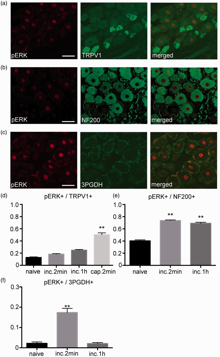
Figure 2.
Distribution of pERK1/2 in the DRG. (a) Double-labeling immunohistochemistry for pERK1/2 and TRPV1 2 min after the incision. A small fraction of the TRPV1 positive neurons expressed pERK1/2 after the incision. Scale bar = 50 µm. (b) Double-labeling immunohistochemistry for pERK1/2 and NF200 at 2 min after the incision. A considerable number of NF200 positive neurons expressed pERK1/2 after the incision. Scale bar = 50 µm. (c) Double-labeling immunohistochemistry for pERK1/2 and 3PGDH 2 min after the incision. pERK1/2 expression was detected in the 3PGDH positive satellite glial cells (SGCs) in the DRG. Scale bar = 50 µm. (d) Although there was no increase in the expression of pERK1/2 in the TRPV1 positive neuron after the incision, there was an increase after the capsaicin injection. n=5 for each group. **: p<0.01 versus the naive group. (e) There was a significant increase in the expression of pERK1/2 in the NF200 positive neuron at 2 min and at 1 h after the incision. n=5 for each group. **: p<0.01 versus the naive group. (f) Fraction of pERK1/2 positive cell area in the 3PGDH positive cell area. There was a significant increase in the pERK1/2 positive area at 2 min after the incision, with a reduction back to baseline values at 1 h after the incision. n=5 for each group. **: p<0.01 versus naive. DRG: dorsal root ganglion; pERK1/2: phosphorylated ERK1/2.

