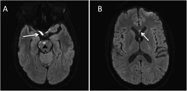
Figure 1.

A, Axial diffusion-weighted magnetic resonance imaging (DW-MRI) demonstrates multifocal areas of restricted diffusion involving the pons (arrowhead) and right prechiasmatic optic nerve (arrow). B, There was additionally restricted diffusion in the corpus callosum (arrow).
