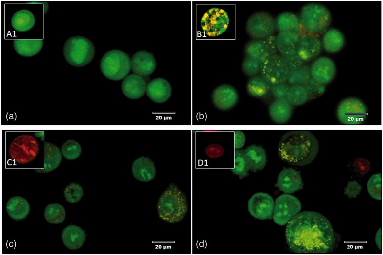
Figure 2.
Morphology and EB/AO staining pattern of apoptotic cells. (a) A normal pneumocyte from the non-treated group with a green-stained appearance and intact nucleus. (b–d) The appearance of the apoptotic pneumocytes showed green to yellow staining with fragmented nuclei in the (b) CPT, (c) HZ, and (d) HZ-IL-1β treatment groups. (A color version of this figure is available in the online journal.)

