Figure 3.
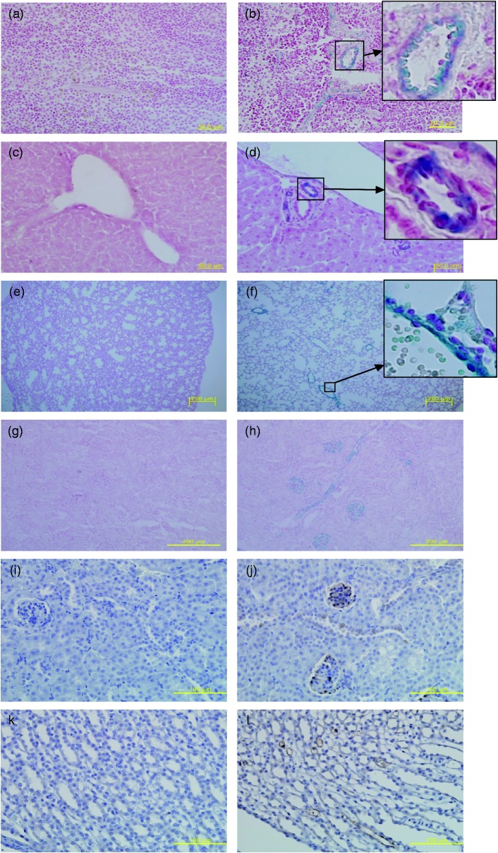
In vivo Fryl gene expression in kidney and other tissues. Frozen mouse tissues were sectioned to 10 µm in thickness and stained with X-gal to detect Fryl gene expression. Nuclear Fast Red was used as counter stain. Tissues from wild (a, c, e, and g) and Fryl+/− (b, d, f, and h) mice were analyzed. Positive staining was found for tubular cells in the spleen (b), liver (d), lung (f), and kidney (h) of Fryl+/−mice. Fryl gene expression in kidneys from nine-week-old wild (i and k) and Fryl+/− (j and l) mice were analyzed by immunohistochemistry staining using anti-β galactosidase antibody. Hematoxylin was used for counterstaining. The glomerulus and collecting tubules showed strong positivity for these staining (j, l). Upper right inlets in b, d, and f show magnified photos of small squares. Magnifications: 400× for (a–d), (i), (j); 100× for e–h; 200× for (k) and (l). (A color version of this figure is available in the online journal.)

