Figure 1.
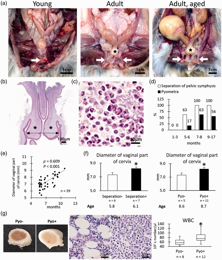
Pathological features of female reproduction system. (a) Gross anatomical features of female cotton rats. Pelvic symphysis is clearly observed in the young group (arrow), but they are separated in the adult group (arrows). Hypertrophy of the vaginal parts of the cervix is also remarkable in the adult group (asterisks). In the adult group, pyometra is evident in some cotton rats. (b) Histology of the vaginal parts of the cervix. Hematoxylin and eosin (HE) staining. Hypertrophy of the lamina propria is observed (asterisks). We measured outer diameters (arrow) of the vaginal parts of the cervix in graph e. (c) Inflammatory cells observed in the lumen of the uterus with pyometra. HE staining. Numerous neutrophils are noted. (d) Incidence of separation of pelvic symphysis and pyometra. (e) The correlation between diameter of the vaginal part of the cervix and age. Spearman’s correlation test (*, P < 0.05). (f) Diameter of the vaginal part of the cervix. Numerical results are expressed as means ± standard errors. Mann–Whitney U test (*, P < 0.05). (g) Gross and histopathology of bone marrow. The bone marrow samples of females with pyometra show more white blood cells, especially neutrophils, than those of females without pyometra. White blood cell (WBC). Box-and-whisker plot. Mann–Whitney U test (*, P < 0.05). (A color version of this figure is available in the online journal.)

