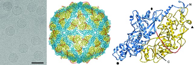
Fig. 1.
Three-dimensional cryo-EM reconstruction of Penicillium chrysogenum virus virions at a resolution of 4.1 Å. (Left) Cryo-EM image of Penicillium chrysogenum virus (scale bar, 50 nm). (Middle) Atomic model of the Penicillium chrysogenum virus capsid viewed along a twofold axis. (Right) Atomic model of a Penicillium chrysogenum virus CP (top view) showing the N-terminal domain (1–498, blue), the linker segment (499–515, red) and the C-terminal domain (516–982, yellow). Symbols indicate icosahedral symmetry axes.

