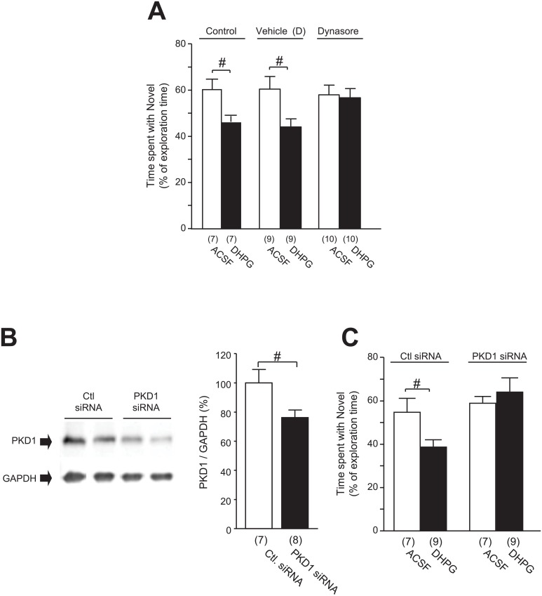
Fig 6. Effects of bilateral infusion of DHPG (50 μmol/2 μL) into the CA1 area on the NOD performance.
The preference for the novel object is represented by the time spent interacting with the novel object as a percentage of the total time spent interacting with objects in A for rats following the infusion of ACSF or DHPG into the CA1 area without (Control, left two bars in A) or with co-application of vehicle of Dynasore (middle two bars in A) or Dynasore (80 μmol, right two bars in A). Blots in B show examples of Western blot analysis of PKD1 (upper blots) and GAPDH proteins (lower blots) in the CA1 area in rats which received the infusion of control siRNA (Ctl. siRNA) or PKD1 siRNA in this area. Summary data of the ratios of band intensities of PKD1 versus GAPDH proteins (= 100%) are shown in the bar graphs in B. Bar graphs in C show the preference for the novel object—represented by the time spent interacting with the novel object as a percentage of the total time spent interacting with objects—of rats following intra-CA1 infusion of ACSF (open bars) or DHPG (filled bars) pre-administrated with PKD1 siRNA or control siRNA. #: p < 0.05 (unpaired t-test); See S5 Table for statistical details; Values in brackets indicate the number of animals tested.

