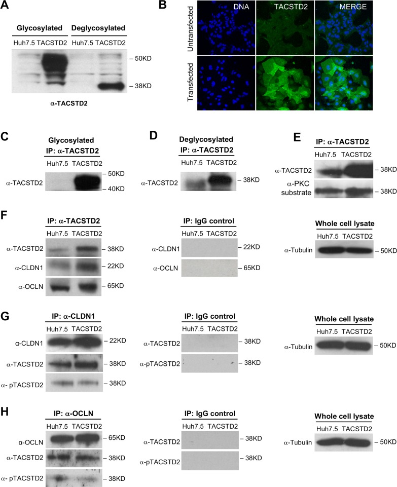
Fig 2. TACSTD2 expression and interaction with HCV-entry cofactors in hepatoma cells.
(A) Western blot analysis of TACSTD2 expression in parental Huh7.5 cells and TACSTD2-overexpressing Huh7.5 cells (TACSTD2). TACSTD2 expression was below the detection limit of Western blot in parental cells (lane 1) but was visible in TACSTD2-overexpressing cells as a broad band due to its poly-glycoslyated state (lane 2). Enzymatic deglycosylation resulted in the protein migration as a single band of 37KD, as seen in lane 4. (B) Immunofluorescence detection of TACSTD2 (green) in parental and TACSTD2-overexpressing Huh7.5 cells. No TACSTD2 could be visualized in parental Huh7.5 (top panel), whereas in stably transfected cells, TACSTD2 was localized both in the cytoplasm and along the cellular membrane (lower panel). (C) Detection of TACSTD2 in parental and TACSTD2-overexpressing Huh7.5 cells by immunoprecipitation with an anti-TACSTD2 antibody without deglycosylation. (D) Detection of TACSTD2 in parental and TACSTD2-overexpressing Huh7.5 cells by immunoprecipitation with an anti-TACSTD2 antibody followed by deglycosylation. (E) Phosphorylation of TACSTD2 in parental and TACSTD2-overexpressing Huh7.5 cells. Cell lysates were immunoprecipitated with anti-TACSTD2 antibody and then deglycosylated. Phosphorylated TACSTD2 was detected using an anti-PKC substrate-specific antibody; total TACSTD2 was detected using biotinylated anti-TACSTD2 antibody. Phosphorylated TACSTD2 was detected in both parental and TACSTD2-overexpressing Huh7.5 cells. (F) Co-immunoprecipitation of CLDN1 and OCLN by anti-TACSTD2 antibody in parental and TACSTD2-overexpressing Huh7.5 cells (lanes 1, 2). Lysates prepared from both parental and TACSTD2-overexpressing Huh7.5 cells were immunoprecipitated with anti-TACSTD2 antibody or control IgG, deglycosylated, and probed with antibodies specific for TACSTD2, CLDN1, and OCLN. (G) Reciprocal co-immunoprecipitation of TACSTD2 by anti-CLDN1 antibody in parental and TACSTD2-overexpressing Huh7.5 cells (lanes 1, 2). Cell lysates were immunoprecipitated with anti-CLDN1 antibody or control IgG. After deglycosylation, total TACSTD2 was detected using biotinylated anti-TACSTD2 antibody, phosphorylated TACSTD2 was detected using an anti-PKC substrate-specific antibody, and CLDN1 was detected using an anti-CLDN1 specific antibody. (H) Reciprocal co-immunoprecipitation of TACSTD2 by anti-OCLN antibody in parental and TACSTD2-overexpressing Huh7.5 cells (lanes 1, 2). Cell lysates were immunoprecipitated with anti-OCLN antibody or control IgG, deglycosylated, and probed with biotinylated anti-TACSTD2 antibody, anti-PKC substrate-specific antibody to detect phosphorylated TACSTD2, and anti-OCLN specific antibody.

