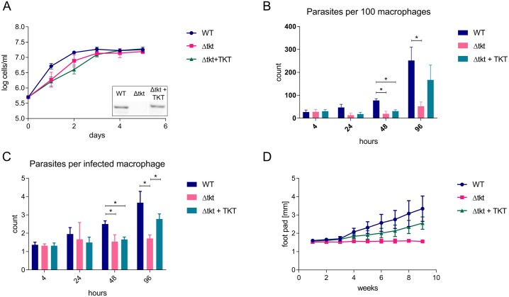
Fig 2. Growth of Δtkt cells in vitro and in vivo.
(A) Growth of WT, Δtkt, and Δtkt + TKT promastigote cells in Homem medium over 5 days, n = 3. The inset shows Western blot analysis of WT, Δtkt, and Δtkt + TKT cell lines probed with α-TKT antibody. (B) Infection of THP-1 macrophages with Leishmania. After 4 hours of incubation the parasites were washed off and infection assessed at time points indicated. 10:1 parasite to host ratio, n = 4, * = p < 0.005. (C) Number of parasites per each infected macrophage was counted, n = 4, * = p < 0.005. (D) Development of foot pad lesions in BALB/c mice infected with WT, Δtkt, and Δtkt + TKT cells, n = 2, four and five mice were used per each group in the two respective replicates.

