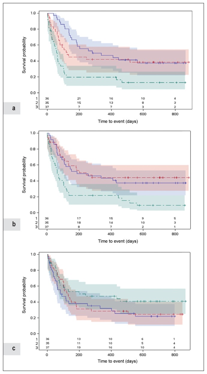
Figure 2.
Progression-free survival according to counts of a) neutrophils, b) platelets, and c) lymphocytes (/μL), Kaiser Permanente Northern California, August 2014 to December 2015.
Follow-up began on the date of initiation of treatment of metastatic melanoma with anti-PD-1 checkpoint inhibitor. Follow-up ended on the earliest date of progression noted on results of imaging or clinical examination (event); or disenrollment from the Health Plan, death of causes other than malignant melanoma, or end of study on February 18, 2017 (censor). We determined the date of progression using the treating oncologist’s assessment guided by imaging studies or clinical symptoms in patients who progressed quickly (within 1–3 cycles of first treatment) before imaging could be performed. Numbers shown at bottom of figures are numbers of patients at risk of progression at each time point based on different range of cell count as indicated (1, blue; 2, red; and 3, green).
PD-1 = programmed cell death protein-1.

