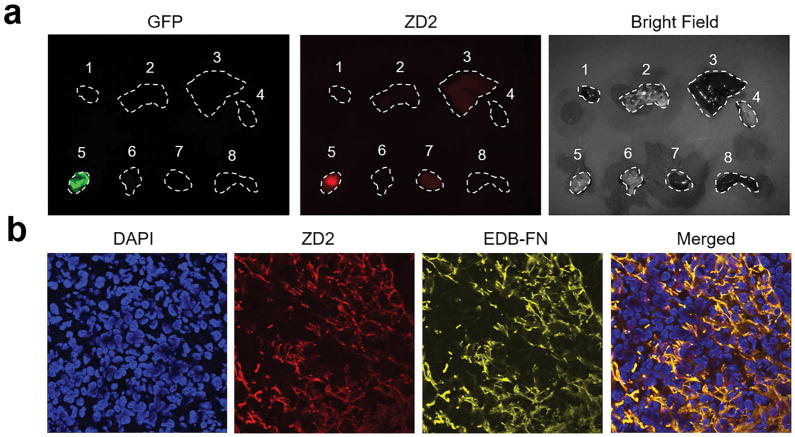
Figure 2. Ex vivo fluorescence imaging and histological analysis of 4T1 tumor.
a. Representative ex vivo fluorescence imaging of tumor and organs harvested from mice bearing 4T1 primary tumor xenografts at 4 h after injection of ZD2-Cy5.5. Numbers denote: 1, heart; 2, lung; 3, liver; 4, muscle; 5, 4T1 tumor; 6, brain; 7, kidney; 8, spleen. b. Histological analysis of the binding of ZD2 peptide and EDB-FN distribution. Merged channel of ZD2 and EDB-FN showed the colocalization of ZD2-Cy5.5 and EDB-FN.

