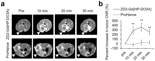
Figure 3. MRI detection of 4T1 primary tumor.
a. Representative axial T1-weighted spin-echo MRI images of 4T1 breast cancer xenografts in mice before and after intravenous injection of ZD2-Gd(HP-DO3A) or Gd(HP-DO3A) at a dose of 0.1 mmol/kg. Tumor locations are denoted with arrowheads. b. CNR analysis of 4T1 tumors (**: P<0.01).

