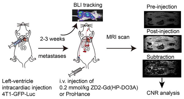
Figure 6. Illustration of 4T1 metastatic tumor model imaging procedures.
Mice were injected with 4T1-GFP-luc cells via the left ventricle of the heart. During 2–3 weeks, BLI was used to track the growth of metastatic tumors throughout the body. MRI was performed before and 20 min after i.v. injection of 0.2 mmol/kg ZD2-Gd(HP-DO3A) or Gd(HP-DO3A). b. Images of muscle, lymph node, and adrenal gland with 4T-GFP-Luc metastatic tumors.

