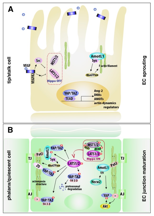
Fig. 1.
Schematic diagram depicting the roles of the Hippo-YAP/TAZ signaling in EC sprouting and junction maturation. (A) VEGF exposure to ECs inactivates LATS1/2, leading to dephosphorylation of AmotL1 and YAP/TAZ in tip and stalk cells. AmotL1 binds with F-actin, regulating the dynamics of F-actin filament via Syx. YAP/TAZ is shuttled into nucleus, increasing expressions of a variety of genes, which are essential for the initiation and expansion of angiogenic sprouting. Syx, Rho GTPase-exchanging factor protein; AmotL1, angiomotin-like 1. (B) Endothelial cell-cell contact induces the Hippo kinase activity. In phalanx cells, LATS1/2 activation phosphorylates AmotL1 and YAP/TAZ. The phosphorylated AmotL1 forms complex with PatJ and Syx to regulate the small Rho GTPase for actomyosin contractility at TJs. The phosphorylated YAP/TAZ leads to the interaction with AmotL1 and ZO proteins at TJs. In addition, the phosphorylated YAP/TAZ is recruited to VE-cadherin-catenin complex at AJs, and degraded in the cytoplasm in the proteasome-dependent proteolysis. Meanwhile, in quiescent ECs, VE-cadherin-mediated Akt activity phosphorylates YAP. AmotL1 is ubiquitinated by Hecw2 and recruited to TJs to promote TJ stability and to seemingly maintain the retention of YAP at EC junction/ cytoplasm. TJ, tight junction; AJ, adherens junction; ZO, zonula occludens; PatJ, polarity-/junction-associated scaffold protein; Hecw2, HECT, C2 and WW domain containing E3 ubiquitin protein ligase 2.

