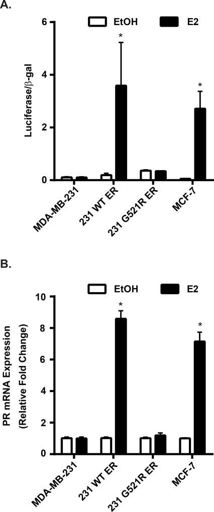
Figure 3. ERα is functionally inactive in 231 G521R ER cells.
(A) Estrogen-deprived cells were transfected with ERE-luciferase and β-galactosidase plasmids and then treated with ethanol (EtOH) vehicle or 10 nM 17β-estradiol (E2) for 24 hours. Luciferase activity was measured and normalized to β-galactosidase activity. (B) PR mRNA expression of estrogen-deprived cells was measured after 24 h treatment with EtOH or 10 nM E2. Values represent the mean±SEM of 3 independent experiments, * p<0.05 compared with the corresponding EtOH control.

