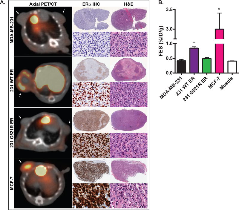
Figure 5. Loss of 18F-FES uptake in 231 G521R ER tumor xenografts.
(A) Representative axial 18F-FES-PET/CT images of tumor xenografts grown in athymic nude female mice. Mice were injected with 250 µCi of 18F-FES and images were obtained 1 hour post injection. Arrows point to tumor. 18F-FES uptake is also noted in gall bladder and liver due to physiologic hepatobiliary clearance. Corresponding low and high power magnification images of ERα immunohistochemistry (IHC) and H&E staining of excised tumors. (B) Quantitative 18F-FES uptake assessed by mean %ID/g. Values represent the mean±SEM, * p<0.05 compared with tumor uptake in MDA-MB-231 and 231 G521R xenografts and compared with non-specific muscle uptake. MDA-MB-231 xenografts (N=8); 231 WT ER xenografts (N=17); 231 G521R ER xenografts (N=9).

