Figure 4. Retinoic acids modulate VEGF secretion by RPE cells.
(A) Choroidal flat-mount immunohistochemistry of E16.5 WT and Aldh1a1–/– embryos stained for endomucin (Emcn, red) and ETS-related gene (ERG, green). Note that hypovascularization and fewer vascular endothelial cells were observed in the dorsal region of Aldh1a1–/– embryonic eyes. (B and C) Quantitative evaluation of the density of Emcn-positive vessels and the number of ERG-positive cells in E16.5 WT and Aldh1a1–/– embryos. Data represent the average ±SD; n = 4 per group. *p<0.05. (D) ELISA analysis of VEGF secreted from WT and Aldh1a1–/– RPE-choroid complex at E17.5 and P24. Data represent the average ±SD; n = 4 independent samples per group. *p<0.05. (E) In situ hybridization on E16.5 WT and Aldh1a1–/– eyes with the Vegfa probe (DIG-labeled, purple). Vegfa expression was reduced in the dorsal RPE cells of Aldh1a1–/– eyes (upper RPEs from red arrowhead). Right panels show the higher magnification images of left panels (four white boxes) and red square brackets indicate RPE layer. (F) Retinoic acid (RA)-dependent enhancement of VEGF secretion of human primary RPE cells evaluated by ELISA. Data are means three times ELISA determinations. ***p<0.001. (G) Choroidal flat-mount immunohistochemistry of P3 WT and Vitamin-A-deficient (VAD) mice stained for endomucin (Emcn, red). Dorsal choroidal hypoplasia was observed in VAD mice. (H) Quantitative evaluation of the vascular density of P3 WT and VAD mice. Data represent the average ±SD; n = 5–6 per group. *p<0.05. (I) Choroidal flat-mount of P3 Aldh1a1–/– and Aldh1a1–/– mice from a mother treated with RA by oral gavage between E10 and E16, immunostained with anti-endomucin antibody (Emcn, red) This RA treatment of the mother restored the choroidal vascularization in P3 Aldh1a1–/– pups. (J) Quantitative evaluation of the vascular density in Aldh1a1–/– mice and Aldh1a1–/– mice from a mother treated with RA. Data represent the average ± SD; n = 4 per group. *p<0.05. N.S., not significant. [Scale bars, 50 μm (A, right panels in E, (G and I), 200 μm (left panels in E)].
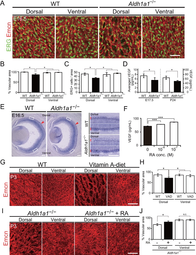
Figure 4—figure supplement 1. Developmental flat-mount immunohistochemical analysis of choroid in the Aldh1a1–/– mice.
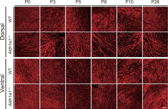
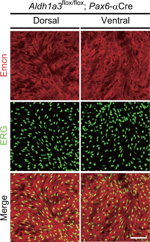
Figure 4—figure supplement 2. Neural retina-specific conditional disruption of Aldh1a3 did not show choroidal hypoplasia in the ventral retina.