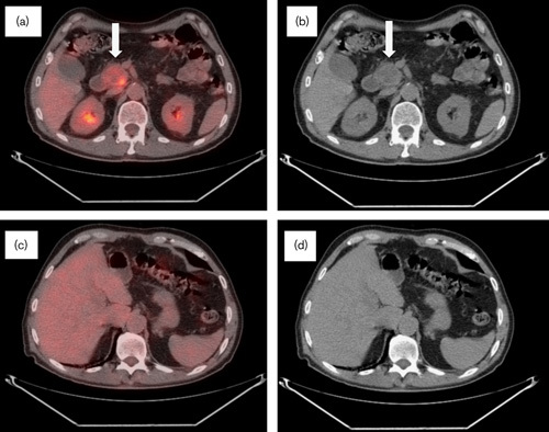
Fig. 4.

CT/PET follow-up performed in November 2008 at 18 months after diagnosis. (a) CT/PET and (b) CT scan: interval decrease in the size of the pancreatic uncinate process mass measuring 3.9×3.6×3.1 cm with increased necrotic components. The metabolic activity showed decreased activity (SUV 6.4). (c) CT/PET and (d) CT scan: no residual metabolic activity and no discrete lesions identified throughout the hepatic parenchyma. No biliary dilatation noted. CT, computed tomography; SUV, standardized uptake value.
